Synovial sarcoma MRI is a critical diagnostic tool for identifying and understanding this rare form of soft tissue cancer. Synovial sarcoma, though uncommon, poses significant challenges due to its aggressive nature and tendency to develop in areas near joints. Magnetic Resonance Imaging (MRI) plays an indispensable role in detecting and characterizing these tumors, providing detailed images that guide treatment decisions. For patients and healthcare providers alike, understanding the importance of MRI in the context of synovial sarcoma is vital for ensuring timely and accurate care.
The journey to diagnosing synovial sarcoma often begins with imaging tests, and MRI is considered the gold standard. Unlike other imaging modalities, MRI offers unparalleled clarity when visualizing soft tissues, making it invaluable for identifying tumors in areas such as the arms, legs, and joints. This imaging technique not only helps confirm the presence of synovial sarcoma but also provides critical information about its size, location, and potential spread. By leveraging MRI, doctors can develop personalized treatment plans tailored to the unique characteristics of each patient’s tumor.
Given the rarity and complexity of synovial sarcoma, staying informed about advancements in MRI technology and interpretation is essential. Modern MRI techniques, such as contrast-enhanced imaging, have significantly improved diagnostic accuracy, enabling earlier detection and better outcomes. As we delve deeper into the topic, we’ll explore the nuances of synovial sarcoma MRI, its role in treatment planning, and how it continues to evolve with advancements in medical science. This article aims to provide a comprehensive guide for patients, caregivers, and healthcare professionals seeking clarity and actionable insights into this critical diagnostic tool.
Read also:Meet The Extraordinary Damian Musk Making Waves In Business And Innovation
Table of Contents
- What is Synovial Sarcoma MRI and Why is it Important?
- How Does Synovial Sarcoma Appear on an MRI Scan?
- Advantages of Using MRI for Diagnosing Synovial Sarcoma
- What Are the Limitations of MRI in Synovial Sarcoma Diagnosis?
- How Can MRI Results Guide Treatment Planning for Synovial Sarcoma?
- Factors Affecting the Accuracy of Synovial Sarcoma MRI
- What Are the Latest Advancements in Synovial Sarcoma MRI Technology?
- Frequently Asked Questions About Synovial Sarcoma MRI
What is Synovial Sarcoma MRI and Why is it Important?
Synovial sarcoma MRI is a specialized imaging technique that uses powerful magnets and radio waves to produce detailed pictures of soft tissues, including tumors. This method is particularly well-suited for detecting synovial sarcoma, a rare type of cancer that typically develops near joints in the arms or legs. Unlike other imaging modalities such as X-rays or CT scans, MRI provides a clearer view of soft tissues, making it an indispensable tool for diagnosing and evaluating this condition.
The importance of synovial sarcoma MRI cannot be overstated. Early and accurate diagnosis is crucial for improving patient outcomes, and MRI plays a pivotal role in achieving this. By offering high-resolution images, MRI helps doctors identify the tumor’s exact location, size, and proximity to critical structures like nerves and blood vessels. This information is essential for determining the most appropriate treatment plan, whether it involves surgery, radiation therapy, or chemotherapy.
Moreover, MRI is non-invasive and does not involve ionizing radiation, making it a safer option for repeated use during follow-up evaluations. This is particularly beneficial for monitoring the effectiveness of treatment or detecting any recurrence of the tumor. In essence, synovial sarcoma MRI serves as both a diagnostic and monitoring tool, empowering healthcare providers to deliver personalized and effective care to patients battling this challenging disease.
How Does Synovial Sarcoma Appear on an MRI Scan?
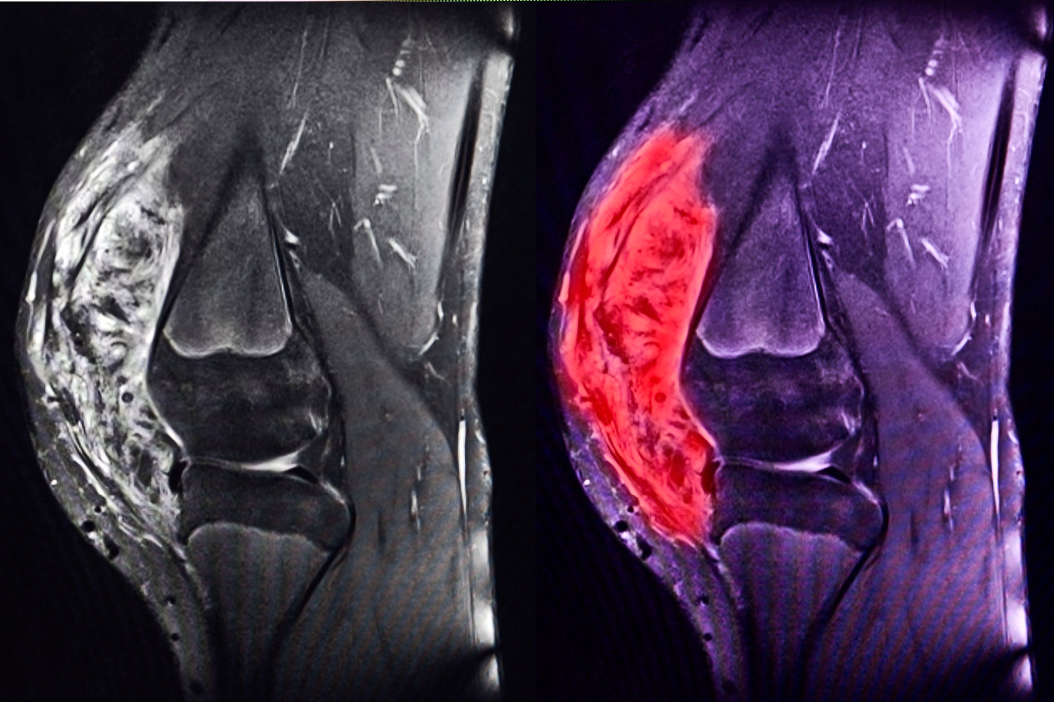
When examining synovial sarcoma MRI results, radiologists look for specific characteristics that differentiate this type of tumor from other soft tissue masses. Typically, synovial sarcoma appears as a well-defined mass with heterogeneous signal intensity on MRI scans. This means that the tumor may exhibit varying shades of brightness depending on the imaging sequence used, such as T1-weighted or T2-weighted images. These variations help highlight the tumor’s composition and its relationship to surrounding tissues.
Key Features of Synovial Sarcoma on MRI
Several hallmark features can help identify synovial sarcoma on MRI:
- Signal Intensity: On T1-weighted images, the tumor often appears isointense or slightly hypointense compared to muscle tissue. On T2-weighted images, it tends to be hyperintense, indicating high water content or fluid within the tumor.
- Contrast Enhancement: When contrast agents are used, synovial sarcoma typically shows heterogeneous enhancement, with some areas lighting up more brightly than others. This pattern reflects the tumor’s varying levels of vascularity and necrosis.
- Calcifications: Synovial sarcoma often contains calcified areas, which appear as dark spots on MRI. These calcifications are a distinguishing feature that aids in diagnosis.
What Do These Features Indicate?
The appearance of synovial sarcoma on MRI provides valuable insights into its nature and behavior. For instance, the presence of calcifications suggests a higher likelihood of malignancy, while the tumor’s heterogeneous signal intensity may indicate areas of necrosis or cystic degeneration. Additionally, the tumor’s proximity to joints, tendons, or neurovascular structures can influence treatment decisions, as these factors may complicate surgical removal or radiation therapy.
Read also:Ultimate Guide To Choosing The Best Support Slippers For Comfort And Health
Interpreting MRI results requires expertise, as synovial sarcoma can sometimes mimic other benign or malignant soft tissue tumors. Radiologists often correlate MRI findings with clinical history, biopsy results, and other imaging studies to arrive at a definitive diagnosis. This multidisciplinary approach ensures that patients receive accurate information and appropriate care tailored to their specific condition.
Advantages of Using MRI for Diagnosing Synovial Sarcoma
MRI stands out as the preferred imaging modality for diagnosing synovial sarcoma due to its numerous advantages over other techniques. Unlike X-rays, which primarily capture bone structures, or CT scans, which provide limited soft tissue detail, MRI offers unparalleled clarity and precision in visualizing soft tissues. This makes it particularly effective for detecting and characterizing tumors like synovial sarcoma, which often develop near joints and other complex anatomical areas.
Superior Soft Tissue Contrast
One of the most significant advantages of MRI is its ability to distinguish between different types of soft tissues. This capability is crucial for identifying synovial sarcoma, as the tumor’s appearance can vary depending on its composition. MRI’s superior contrast resolution allows radiologists to differentiate between tumor tissue, muscle, fat, and other structures, providing a comprehensive view of the lesion’s characteristics. This level of detail is invaluable for accurate diagnosis and treatment planning.
Non-Invasive and Safe
Another key benefit of MRI is its non-invasive nature. Unlike procedures that involve ionizing radiation, such as CT scans or X-rays, MRI uses powerful magnets and radio waves to generate images. This makes it a safer option for repeated use, especially during follow-up evaluations to monitor treatment effectiveness or detect recurrence. Patients undergoing MRI experience no exposure to harmful radiation, reducing the risk of long-term side effects.
Customizable Imaging Protocols
MRI also offers the flexibility to tailor imaging protocols to the specific needs of each patient. For instance, contrast-enhanced MRI can highlight areas of increased vascularity within the tumor, providing additional information about its aggressiveness. Similarly, specialized sequences like diffusion-weighted imaging (DWI) can help assess the tumor’s cellular density, offering insights into its biological behavior. This adaptability ensures that MRI remains a versatile and indispensable tool in the diagnosis and management of synovial sarcoma.
What Are the Limitations of MRI in Synovial Sarcoma Diagnosis?
While MRI is an invaluable tool for diagnosing synovial sarcoma, it is not without its limitations. Understanding these constraints is essential for interpreting MRI results accurately and ensuring that patients receive comprehensive care. One of the primary challenges lies in the potential for misinterpretation, as synovial sarcoma can sometimes mimic other soft tissue tumors or benign lesions on MRI scans.
Challenges in Differentiating Synovial Sarcoma from Other Tumors
Synovial sarcoma often shares imaging characteristics with other soft tissue masses, such as liposarcomas, fibrosarcomas, or even benign cysts. For example, the heterogeneous signal intensity and contrast enhancement patterns seen in synovial sarcoma can also appear in other types of tumors. This overlap can make it difficult for radiologists to arrive at a definitive diagnosis based solely on MRI findings, necessitating additional tests like biopsies or PET scans to confirm the nature of the lesion.
Limitations in Assessing Tumor Spread
Another limitation of MRI is its reduced effectiveness in detecting distant metastases, particularly in areas like the lungs or bones. While MRI excels at visualizing soft tissues, it may not provide the same level of clarity when assessing tumors in organs with different compositions. In such cases, complementary imaging modalities like CT scans or PET-CT may be required to evaluate the full extent of the disease. This underscores the importance of a multidisciplinary approach to diagnosis and treatment planning.
What Can Be Done to Overcome These Limitations?
To mitigate these limitations, healthcare providers often combine MRI with other diagnostic tools and clinical information. For instance, correlating MRI findings with biopsy results and patient history can help reduce the risk of misdiagnosis. Additionally, advancements in MRI technology, such as the use of artificial intelligence algorithms, are being explored to improve the accuracy and reliability of imaging interpretations. These innovations hold promise for enhancing the diagnostic capabilities of MRI in the context of synovial sarcoma.
How Can MRI Results Guide Treatment Planning for Synovial Sarcoma?
MRI results play a pivotal role in shaping the treatment strategy for synovial sarcoma, offering critical insights that inform every step of the process. From determining the feasibility of surgery to planning radiation therapy, the detailed images provided by MRI enable healthcare providers to develop personalized and effective treatment plans tailored to each patient’s unique condition.
Assessing Surgical Feasibility
One of the primary ways MRI results guide treatment planning is by assessing whether surgical removal of the tumor is feasible. The high-resolution images produced by MRI allow surgeons to evaluate the tumor’s size, location, and proximity to critical structures such as nerves, blood vessels, and joints. This information is crucial for determining whether complete resection is possible without causing significant functional impairment or complications. In cases where the tumor is deeply embedded or intertwined with vital structures, MRI findings may prompt a discussion about alternative treatment options, such as radiation or chemotherapy.
Planning Radiation Therapy
MRI also plays a key role in planning radiation therapy for synovial sarcoma. By providing detailed images of the tumor and surrounding tissues, MRI helps radiation oncologists precisely target the lesion while minimizing exposure to healthy tissues. This precision is particularly important for tumors located near sensitive areas, such as the spine or major joints, where excessive radiation could lead to long-term complications. Additionally, MRI can help monitor the tumor’s response to radiation therapy over time, allowing for adjustments to the treatment plan as needed.
What Role Does MRI Play in Follow-Up Evaluations?
After treatment, MRI remains an essential tool for monitoring the patient’s progress and detecting any signs of recurrence. Regular follow-up scans can help identify residual tumor tissue or new lesions early, enabling timely intervention. This ongoing surveillance is critical for ensuring the best possible outcomes and maintaining the patient’s quality of life. By leveraging MRI throughout the treatment journey, healthcare providers can deliver comprehensive and adaptive care that addresses the evolving needs of patients with synovial sarcoma.
Factors Affecting the Accuracy of Synovial Sarcoma MRI
The accuracy of synovial